First of all, We have to know what is Memory?
When an event happens, when you learn something, or when you meet someone, your brain determines whether that information needs to be saved. If your brain judges the information important, it places it in your memory as like "files." You probably know your brain has different analitical parts. Some of them are important for memory. The hippocampus (say: hih-puh-kam-pus) is one of the more important parts of the brain that processes memories. Old information and new information, or memories, are thought to be processed and stored away in different areas of the cerebral cortex, or the "gray matter" of the brain - the largest, outermost part of the brain.
Let us know more about hippocampus.
The hippocampus is a part of the brain located inside the temporal lobe (humans and other mammals have two hippocampi, one in each side of the brain). It forms a part of the limbic system and plays a part in memory and spatial navigation. The name derives from its curved shape in coronal sections of the brain, which resembles a seahorse (Greek: hippo=horse, kampos=sea monster). In Alzheimer's disease, the hippocampus becomes one of the first regions of the brain to suffer damage; memory problems and disorientation appear amongst the first symptoms. Damage to the hippocampus can also result from oxygen starvation (anoxia) and encephalitis. In the anatomy of animals, the hippocampus is among the phylogenetically oldest parts of the brain. The hippocampal emergence from the archipallium is most pronounced in primates and Cetacean sea mammals. Nonetheless, in primates the hippocampus occupies less of the cerebrum in proportion to cerebral cortex among the youngest species, especially humans. The significant development of hippocampal volume in primates correlates more with overall increase of brain mass than with neocortical development.
ANATOMY OF HIPPOCAMPUS
Although there is a lack of consensus relating to terms describing the hippocampus and the adjacent cortex, the term hippocampal formation generally applies to the dentate gyrus, the Cornu Ammonis fields CA1-CA3 (and CA4, frequently called the hilus and considered part of the dentate gyrus), and the subiculum. The CA1, CA2 and CA3 fields make up the hippocampus proper.
Information flow through the hippocampus proceeds from dentate gyrus to CA3 to CA1 to the subiculum, with additional input information at each stage and outputs at each of the two final stages. CA2 represents only a very small portion of the hippocampus and its presence is often ignored in accounts of hippocampal function, though it is notable that this small region seems unusually resistant to conditions that usually cause large amounts of cellular damage, such as epilepsy.
The perforant path, which brings information primarily from entorhinal cortex (but also perirhinal cortex, among others), is generally considered the main source of input to the hippocampus. Layer II of entorhinal cortex (EC) brings input to the dentate gyrus and field CA3, while EC layer III brings input to field CA1 and the subiculum. The main output pathways of the hippocampus are the cingulum bundle and the fimbria/fornix, which arise from field CA1 and the subiculum.

Diagram of hippocampus
Perforant path input from EC layer II enters the dentate gyrus and is relayed to region CA3 (and to mossy cells, located in the hilus of the dentate gyrus, which then send information to distant portions of the dentate gyrus where the cycle is repeated). Region CA3 combines this input with signals from EC layer II and sends extensive connections within the region and also sends connections to region CA1 through a set of fibers called the Schaffer collaterals. Region CA1 receives input from the CA3 subfield, EC layer III and the nucleus reuniens of the thalamus (which project only to the terminal apical dendritic tufts in the stratum lacunosum-moleculare). In turn, CA1 projects to the subiculum as well as sending information along the aforementioned output paths of the hippocampus. The subiculum is the final stage in the pathway, combining information from the CA1 projection and EC layer III to also send information along the output pathways of the hippocampus.
The hippocampus also receives a number of subcortical inputs. In Macaca fascicularis, these inputs include the amygdala (specifically the anterior amygdaloid area, the basolateral nucleus, and the periamygdaloid cortex), the medial septum and the diagonal band of Broca, the claustrum, the substantia innominata and the basal nucleus of Meynert, the thalamus (including the anterior nuclear complex, the laterodorsal nucleus, the paraventricular and parataenial nuclei, the nucleus reuniens, and the nucleus centralis medialis), the lateral preoptic and lateral hypothalamic areas, the supramammillary and retromammillary regions, the ventral tegmental area, the tegmental reticular fields, the raphe nuclei (the nucleus centralis superior and the dorsal raphe nucleus), the nucleus reticularis tegementi pontis, the central gray, the dorsal tegmental nucleus, and the locus coeruleus.
It is widely accepted that each of these regions has a unique functional role in the information processing of the hippocampus, but to date the specific contribution of each region is poorly understood.
Information flow through the hippocampus proceeds from dentate gyrus to CA3 to CA1 to the subiculum, with additional input information at each stage and outputs at each of the two final stages. CA2 represents only a very small portion of the hippocampus and its presence is often ignored in accounts of hippocampal function, though it is notable that this small region seems unusually resistant to conditions that usually cause large amounts of cellular damage, such as epilepsy.
The perforant path, which brings information primarily from entorhinal cortex (but also perirhinal cortex, among others), is generally considered the main source of input to the hippocampus. Layer II of entorhinal cortex (EC) brings input to the dentate gyrus and field CA3, while EC layer III brings input to field CA1 and the subiculum. The main output pathways of the hippocampus are the cingulum bundle and the fimbria/fornix, which arise from field CA1 and the subiculum.
Diagram of hippocampus
Perforant path input from EC layer II enters the dentate gyrus and is relayed to region CA3 (and to mossy cells, located in the hilus of the dentate gyrus, which then send information to distant portions of the dentate gyrus where the cycle is repeated). Region CA3 combines this input with signals from EC layer II and sends extensive connections within the region and also sends connections to region CA1 through a set of fibers called the Schaffer collaterals. Region CA1 receives input from the CA3 subfield, EC layer III and the nucleus reuniens of the thalamus (which project only to the terminal apical dendritic tufts in the stratum lacunosum-moleculare). In turn, CA1 projects to the subiculum as well as sending information along the aforementioned output paths of the hippocampus. The subiculum is the final stage in the pathway, combining information from the CA1 projection and EC layer III to also send information along the output pathways of the hippocampus.
The hippocampus also receives a number of subcortical inputs. In Macaca fascicularis, these inputs include the amygdala (specifically the anterior amygdaloid area, the basolateral nucleus, and the periamygdaloid cortex), the medial septum and the diagonal band of Broca, the claustrum, the substantia innominata and the basal nucleus of Meynert, the thalamus (including the anterior nuclear complex, the laterodorsal nucleus, the paraventricular and parataenial nuclei, the nucleus reuniens, and the nucleus centralis medialis), the lateral preoptic and lateral hypothalamic areas, the supramammillary and retromammillary regions, the ventral tegmental area, the tegmental reticular fields, the raphe nuclei (the nucleus centralis superior and the dorsal raphe nucleus), the nucleus reticularis tegementi pontis, the central gray, the dorsal tegmental nucleus, and the locus coeruleus.
It is widely accepted that each of these regions has a unique functional role in the information processing of the hippocampus, but to date the specific contribution of each region is poorly understood.
Role of Hippocampus in General Memory
Drawing of the neural circuitry of the rodent hippocampus. S. Ramón y Cajal, 1911.Psychologists and neuroscientists dispute the precise role of the hippocampus, but, in general, agree that it has an essential role in the formation of new memories about experienced events (episodic or autobiographical memory). Some researchers prefer to consider the hippocampus as part of a larger medial temporal lobe memory system responsible for general declarative memory (memories that can be explicitly verbalized — these would include, for example, memory for facts in addition to episodic memory). Some evidence supports the idea that, although these 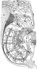 forms of memory often last a lifetime, the hippocampus ceases to play a crucial role in the retention of the memory after a period of consolidation. Damage to the hippocampus usually results in profound difficulties in forming new memories (anterograde amnesia), and normally also affects access to memories prior to the damage (retrograde amnesia). Although the retrograde effect normally extends some years prior to the brain damage, in some cases older memories remain - this sparing of older memories leads to the idea that consolidation over time involves the transfer of memories out of the hippocampus to other parts of the brain. However, experimentation has difficulties in testing the sparing of older memories; and, in some cases of retrograde amnesia, the sparing appears to affect memories formed decades before the damage to the hippocampus occurred, so its role in maintaining these older memories remains controversial. Damage to the hippocampus does not affect some aspects of memory, such as the ability to learn new skills (playing a musical instrument, for example), suggesting that such abilities depend on a different type of memory (procedural memory) and different brain regions. And there is evidence (e.g., O'Kane et al 2004) to suggest that patient HM (who had his medial temporal lobes removed bilaterally as a treatment for epilepsy) can form new semantic memories.
 forms of memory often last a lifetime, the hippocampus ceases to play a crucial role in the retention of the memory after a period of consolidation. Damage to the hippocampus usually results in profound difficulties in forming new memories (anterograde amnesia), and normally also affects access to memories prior to the damage (retrograde amnesia). Although the retrograde effect normally extends some years prior to the brain damage, in some cases older memories remain - this sparing of older memories leads to the idea that consolidation over time involves the transfer of memories out of the hippocampus to other parts of the brain. However, experimentation has difficulties in testing the sparing of older memories; and, in some cases of retrograde amnesia, the sparing appears to affect memories formed decades before the damage to the hippocampus occurred, so its role in maintaining these older memories remains controversial. Damage to the hippocampus does not affect some aspects of memory, such as the ability to learn new skills (playing a musical instrument, for example), suggesting that such abilities depend on a different type of memory (procedural memory) and different brain regions. And there is evidence (e.g., O'Kane et al 2004) to suggest that patient HM (who had his medial temporal lobes removed bilaterally as a treatment for epilepsy) can form new semantic memories.
 Role of Hippocampus in spatial memory and navigation
Some evidence implicates the hippocampus in storing and processing spatial information. Studies in rats have shown that neurons in the hippocampus have spatial firing fields. These cells are called place cells. Some cells fire when the animal finds itself in a particular location, regardless of direction of travel, while most are at least partially sensitive to head direction and direction of travel. In rats, some cells, termed context-dependent cells, may alter their firing depending on the animal's recent past (retrospective) or expected future (prospective). Different cells fire at different locations, so that, by looking at the firing of the cells alone, it becomes possible to tell where the animal is. Place cells have now been seen in humans involved in finding their way around in a virtual reality town. The findings resulted from research with individuals that had electrodes implanted in their brains as a diagnostic part of surgical treatment for serious epilepsy. The discovery of place cells led to the idea that the hippocampus might act as a cognitive map — a neural representation of the layout of the environment. Recent evidence has cast doubt on this perspective, indicating that the hippocampus might be crucial for more fundamental processes within navigation. Regardless, studies with animals have shown that an intact hippocampus is required for simple spatial memory tasks (for instance, finding the way back to a hidden goal). Without a fully-functional hippocampus, humans may not successfully remember where they have been and how to get where they are going. Researchers believe that the hippocampus plays a particularly important role in finding shortcuts and new routes between familiar places. Some people exhibit more skill at this sort of navigation than do others, and brain imaging shows that these individuals have more active hippocampi when navigating. London's taxi drivers must learn a large number of places — and know the most direct routes between them (they have to pass a strict test, The Knowledge, before being licensed to drive the famous black cabs). A study at University College London (Maguire et al, 2000) showed that part of the hippocampus is larger in taxi drivers than in the general public, and that more-experienced drivers have bigger hippocampi. Whether having a bigger hippocampus helps an individual to become a cab driver or finding shortcuts for a living makes an individual's hippocampus grow is yet to be elucidated. A study on rats at Indiana University suggested that the sexual dimorphism in the hippocampus morphology is tied to a sexual dimorphism in repeated maze performance. Males seem to be better at contexualizing their whereabouts because they have more hippocampus to work with.
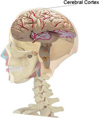
Now let us know more about cerebral cortex
The cerebral cortex is a brain structure in vertebrates. In non-living, preserved brains, the outermost layers of the cerebrum has a grey color, hence the name "grey matter". Grey matter is formed by neurons and their unmyelinated fibers while the white matter below the grey matter of the cortex is formed predominantly by myelinated axons interconnecting different  regions of the central nervous system. The human cerebral cortex is 2-4 mm (0.08-0.16 inches) thick and plays a central role in many complex brain functions including memory, attention, perceptual awareness, "thinking", language and consciousness. The surface of the cerebral cortex is folded in large mammals like humans, where more than two thirds of the cortical surface is buried in the grooves, called "sulci". The phylogenetically more ancient part of the cerebral cortex, the hippocampus, is differentiated in five layers, while the more recent neo-cortex is differentiated in six basic layers. Relative variations in thickness or cell type (among other parametres) allows us to distinguish among different neocortical architectonic fields. The geometry of these fields seems to be related to the anatomy of the cortical folds and, for example, layers in the upper part of the cortical grooves (called gyri) are more clearly differentiated than in its deeper parts (called sulcal "fundi").
 regions of the central nervous system. The human cerebral cortex is 2-4 mm (0.08-0.16 inches) thick and plays a central role in many complex brain functions including memory, attention, perceptual awareness, "thinking", language and consciousness. The surface of the cerebral cortex is folded in large mammals like humans, where more than two thirds of the cortical surface is buried in the grooves, called "sulci". The phylogenetically more ancient part of the cerebral cortex, the hippocampus, is differentiated in five layers, while the more recent neo-cortex is differentiated in six basic layers. Relative variations in thickness or cell type (among other parametres) allows us to distinguish among different neocortical architectonic fields. The geometry of these fields seems to be related to the anatomy of the cortical folds and, for example, layers in the upper part of the cortical grooves (called gyri) are more clearly differentiated than in its deeper parts (called sulcal "fundi").
 Now what can go wrong with Memory?
As wonderful as memory is, it isn't always perfect. It's normal to occasionally forget the name of somebody you just met or where you put your shoes[U said Chromosome, may be]. And of course, everyone has forgotten an answer on a test. Darn! You knew that one, too! It's also typical for people to forget more things as they grow older. Your parents or grandparents might joke about having a "senior moment." That's when they forget something. But some memory problems are serious, such as when a person has Alzheimer's disease. Strokes, which also affect older people, are another medical problem that can affect someone's memory. A stroke is when the blood supply to the brain is temporarily stopped or when a blood vessel bursts. Another problem could occuer, that is TBI or Traumatic Brain Injury which can impair your memory for forever.


.jpg)
2 comments:
this piece of information about memory is really nice!
keep it up!
THANX!
Post a Comment